

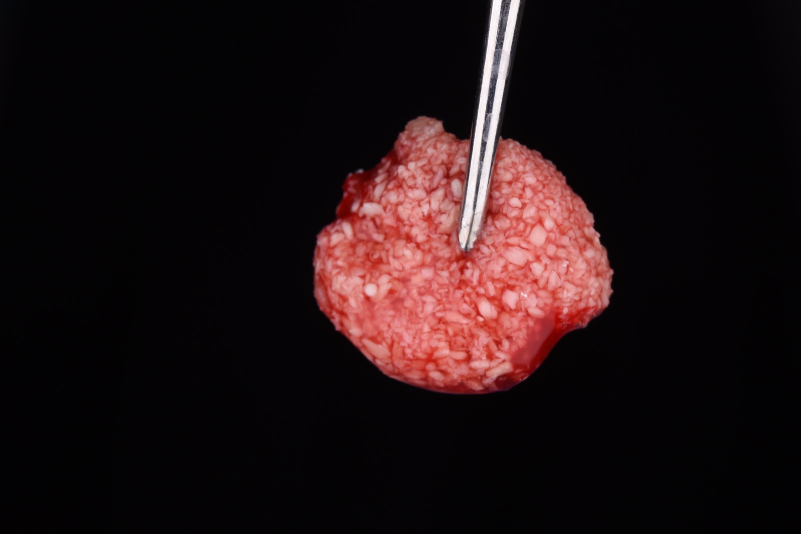
Sticky bone is one of the highest-value, lowest-cost upgrades you can bring into a surgical practice: a moldable, autologous graft that holds its shape, resists being washed out, and carries your patient's own growth factors into the site. The biology is proven; what trips people up is the how. This issue is a build-it-in-your-office guide — get the spin right, make a clean PRF membrane, and assemble sticky bone the way it's meant to be made.
What You'll Need
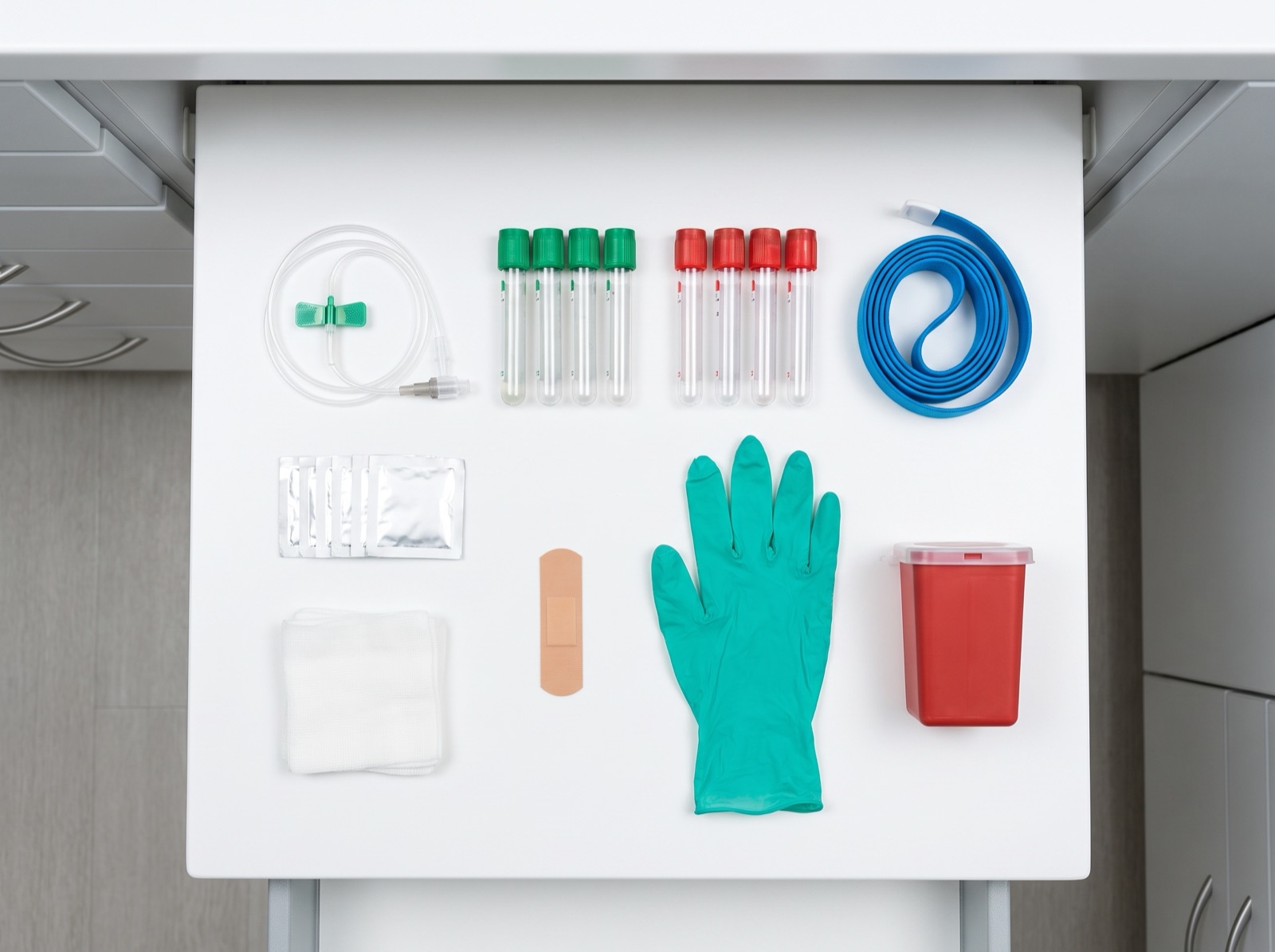
Step 1 — Spin It Right (g-Force, Not RPM)
The single most common error is dialing RPM. RPM only tells you how fast the rotor turns; the force the cells actually feel — relative centrifugal force (RCF, or g-force) — also depends on your rotor's radius. The same RPM on two machines delivers different g-forces. Set g-force if your centrifuge allows it (most modern units do).
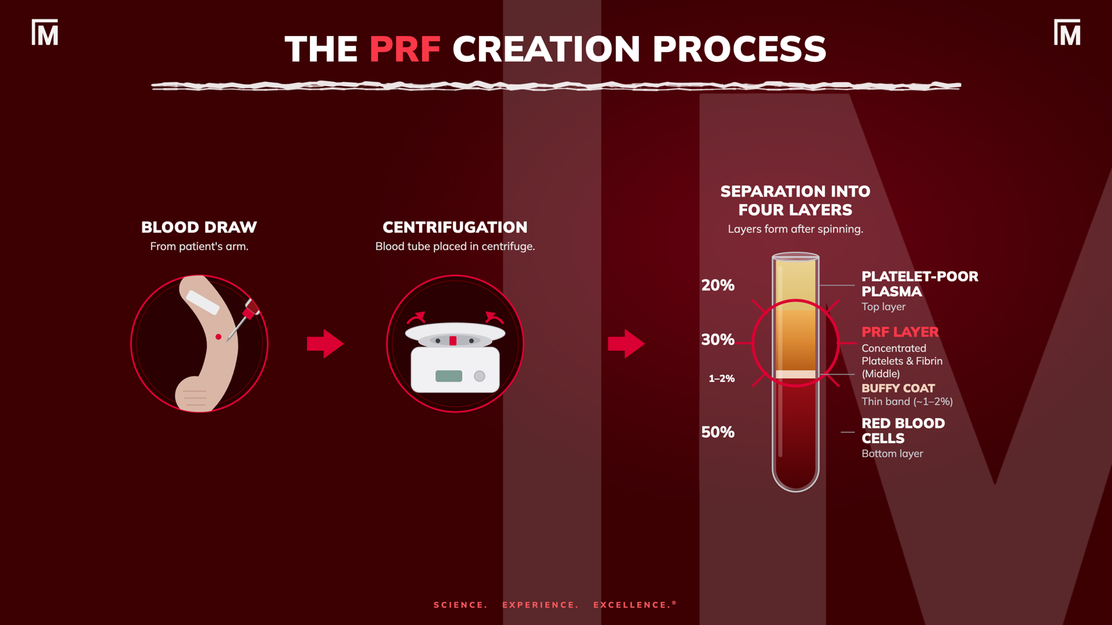
Target These g-Forces
| Product | Set g-force | Time | Tube(s) |
|---|---|---|---|
| PRF membrane (solid) | 700 g | 8 min | Red / glass |
| Sticky bone (co-spin) | 700 g | 8 min | Red / glass + white/blue PET, together |
| A-PRF+ membrane (softer alt.) | 200 g | 8 min | Red / glass |
| i-PRF (injectable, low-speed) | 60 g | 3 min | White/blue PET |
| C-PRF (max concentration liquid) | 2000 g | 8 min | White/blue PET; harvest buffy coat |
For sticky bone, everything spins at one setting — 700 g for 8 min — with the liquid (white/blue) and solid (red) tubes in the rotor together.
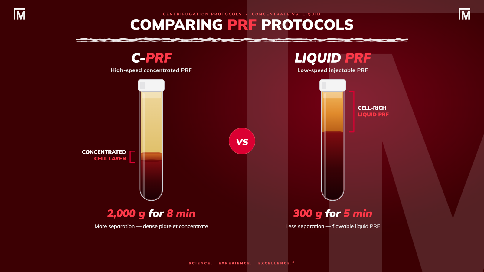
If your machine only shows RPM — convert with RPM = 1000 × √( RCF ÷ (11.18 × r) ), where r is your rotor radius in cm. On a horizontal rotor measure to the tube tip swung out; on a fixed-angle rotor measure to the lowest outer corner of the tilted tube. Common clinical rotors (Scilogex DM0412, Choukroun Duo) are ~11 cm.
| Rotor radius | 700 g (membrane / sticky) | 200 g (A-PRF+) | 60 g (i-PRF) | 2000 g (C-PRF) |
|---|---|---|---|---|
| 9.5 cm | 2,570 rpm | 1,375 rpm | 750 rpm | 4,340 rpm |
| 11 cm (DM0412 / Duo) | 2,400 rpm | 1,275 rpm | 700 rpm | 4,035 rpm |
| 12.7 cm | 2,220 rpm | 1,190 rpm | 650 rpm | 3,755 rpm |
| 14 cm | 2,115 rpm | 1,130 rpm | 620 rpm | 3,575 rpm |
Same formula for horizontal and fixed-angle — only the radius measurement differs. Round to your dial; confirm against your rotor.
Step 2 — Make a PRF Membrane
- Draw blood into red / glass tubes.
- Spin 700 g × 8 min (or 200 g × 8 min for a softer, more cell-distributed A-PRF+ membrane).
- Remove the lid and wait ~5 min — this lets the clot mature and retract so it releases cleanly.
- Lift the clot out; gently separate the red-cell tail with scissors.
- Place in the PRF box, close, and compress 2–5 min.
- Membrane ready — use as a barrier / wound cover, or cut into fragments for sticky bone (see next).
Step 3 — Make Sticky Bone
The reliable recipe uses both components: chopped L-PRF membranes for fibrin scaffold and body, and liquid PRF as the binder that clots the graft into a cohesive mass.
- At the draw, fill the white/blue (liquid) tube FIRST, then the red tubes (see pearl below).
- Spin 700 g × 8 min with 2 white/blue tubes + 2–4 red tubes together.
- Remove the lids of the red tubes; wait 5 min.
- Remove the clots from the red tubes.
- Place clots in the PRF box and allow to compress into membranes for 2–5 min.
- Cut up ~2 membranes (into many small pieces) and place in the PRF bowl — mix with allograft — this is your bone-graft complex.
- With an 18 G × 1.5″ needle, draw the liquid-PRF from the white/blue tubes.
- Mix the liquid-PRF into the bone-graft complex and let it set a few seconds.
- Sticky bone is ready to shape and place.
Fixed-angle note: co-spin at 700 g × 8 min works, but angled rotors nudge the liquid toward clotting — draw the liquid within ~10–15 min of the spin. If a PET tube has started to gel, next time pull the liquid tubes ~5 min into the spin and hold them while you finish the membranes.
The Pearls That Make It Work Every Time
"Draw first, add last." These sound opposite but describe two different moments.
Draw first — at phlebotomy, fill the liquid (white/blue) tube first; the freshest, least-activated blood makes the best liquid PRF.
Add last — during assembly, build the membrane-and-allograft complex first, then draw the liquid and mix it in as the final step, because it sets within seconds of touching the graft. (That's why, in Miron's demo, the liquid isn't drawn until after the membranes are chopped and mixed.)
• Want it firmer? Warm the mix toward ~37 °C (even briefly) to set faster and denser, or give it 10–15 min to fully polymerize. More membrane fragments = more body.
• Keep it clean. Additive-free tubes only; balance the rotor; use everything promptly once the liquid is drawn.
Does It Hold Up? (The Honest Version)
Sticky bone gives you handling and graft stability — it resists being washed out and keeps particles where you place them. Systematic reviews support PRF as a bone-graft adjunct for ridge preservation, intrabony defects, GBR, and sinus grafting (Miron et al., 2021; Fujioka-Kobayashi et al., 2021), and the AAP's 2022 network meta-analysis found adding a biologic to a graft improves clinical and radiographic outcomes over graft or flap alone (Tavelli et al., 2022). Generally, we see improved histological outcomes (more blood vessels in vital bone vs. graft particles) in bone grafts, and improved patient reported pain experience, however its can't do everything.
Clinicians using a PRF membrane in place of a collagen membrane are missing the mark. It doesn't provide any cell exclusion, and you're not "saving money on your membrane" by using it in this way. However, if you're looking for something that can improve handeling during surgery, improve vital bone formation and early healing markers, then you're in the right place.
Keep expectations calibrated: for example, in root coverage, PRF does not replace a connective tissue graft (Chambrone et al., 2022). Think of sticky bone as a better-handling, growth-factor-enriched graft — not a different biologic class.
FROM THE LITERATURE
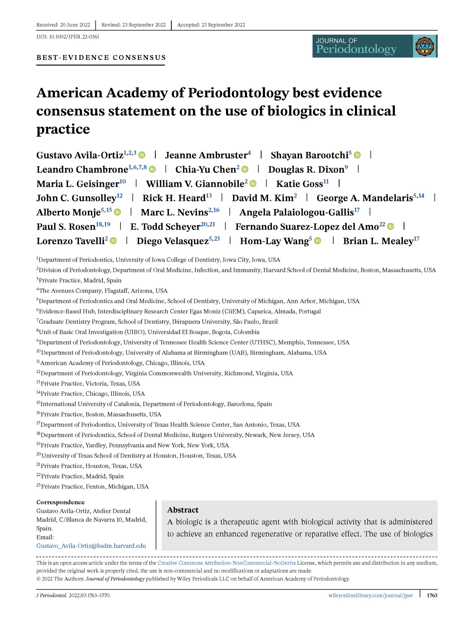
American Academy of Periodontology best evidence consensus statement on the use of biologics in clinical practice
Avila-Ortiz G, et al. J Periodontol. 2022;93:1763–1770. The umbrella consensus behind the Tavelli and Chambrone network meta-analyses cited here.
START MONDAY!
- Set your centrifuge to 700 g / 8 min and co-spin red + white/blue tubes for sticky bone.
- Fill the liquid tube first at the draw; add the liquid last at assembly.
- Chop 2 membranes into the allograft, then bind with liquid PRF — membranes and liquid, not liquid alone.
MORE FROM MISCH IMPLANT IQ
All issues: Misch Implant IQ archive
UPCOMING MISCH INSTITUTE COURSES
Fresh dates are posted as they're announced — see the full institute calendar at misch.com.
Full calendar and registration at misch.com.
Sources
1. Miron RJ, Fujioka-Kobayashi M, Sculean A, Zhang Y. Optimization of platelet-rich fibrin. Periodontol 2000. 2024;94(1):79–91. · PubMed · DOI
2. Miron RJ, Chai J, Fujioka-Kobayashi M, Sculean A, Zhang Y. Evaluation of 24 protocols for the production of platelet-rich fibrin. BMC Oral Health. 2020;20(1):310. · PubMed · DOI
3. Miron RJ, Chai J, Zheng S, et al. A novel method for evaluating and quantifying cell types in PRF and an introduction to horizontal centrifugation. J Biomed Mater Res A. 2019;107(10):2257–2271. · PubMed · DOI
4. Miron RJ, Fujioka-Kobayashi M, Moraschini V, et al. Efficacy of PRF on bone formation, part 1: alveolar ridge preservation. Int J Oral Implantol (Berl). 2021;14(2):181–194.
5. Fujioka-Kobayashi M, Miron RJ, Moraschini V, et al. Efficacy of PRF on bone formation, part 2: GBR, sinus elevation and implant therapy. Int J Oral Implantol (Berl). 2021;14(3):285–302.
6. Tavelli L, Chen C-YJ, Barootchi S, Kim DM. Efficacy of biologics for the treatment of periodontal infrabony defects: an AAP best evidence systematic review and network meta-analysis. J Periodontol. 2022;93(12):1803–1826. · PubMed · DOI
7. Chambrone L, Barootchi S, Avila-Ortiz G. Efficacy of biologics in root coverage and gingival augmentation therapy: an AAP best evidence systematic review and network meta-analysis. J Periodontol. 2022;93(12):1771–1802. · PubMed · DOI
Tsujino T, Takahashi A, Yamaguchi S, et al.. Evidence for contamination of silica microparticles in advanced platelet-rich fibrin matrices prepared using silica-coated plastic tubes. Biomedicines. 2019;7(2):45. · PubMed · DOI
Fujioka-Kobayashi M, Miron RJ, Hernandez M, Kandalam U, Zhang Y, Choukroun J. Optimized platelet-rich fibrin with the low-speed concept: growth factor release, biocompatibility, and cellular response. J Periodontol. 2017;88(1):112-121. · PubMed · DOI
Miron RJ, Fujioka-Kobayashi M, Hernandez M, et al.. Injectable platelet rich fibrin (i-PRF): opportunities in regenerative dentistry?. Clin Oral Investig. 2017;21(8):2619-2627. · PubMed · DOI
Miron RJ, Chai J, Zhang P, et al.. A novel method for harvesting concentrated platelet-rich fibrin (C-PRF) with a 10-fold increase in platelet and leukocyte yields. Clin Oral Investig. 2020;24(8):2819-2828. · PubMed · DOI
Miron RJ, Fujioka-Kobayashi M, Moraschini V, Zhang Y, Gruber R, Wang HL. Efficacy of platelet-rich fibrin on bone formation, part 1: alveolar ridge preservation. Int J Oral Implantol (Berl). 2021;14(2):181-194. · PubMed
Fujioka-Kobayashi M, Miron RJ, Moraschini V, Zhang Y, Gruber R, Wang HL. Efficacy of platelet-rich fibrin on bone formation, part 2: guided bone regeneration, sinus elevation and implant therapy. Int J Oral Implantol (Berl). 2021;14(3):285-302. · PubMed
Avila-Ortiz G, Ambruster J, Barootchi S, et al.. American Academy of Periodontology best evidence consensus statement on the use of biologics in clinical practice. J Periodontol. 2022;93(12):1763-1770. · PubMed · DOI
Misch Implant IQ · Educational content — not a substitute for individual clinical judgment.