

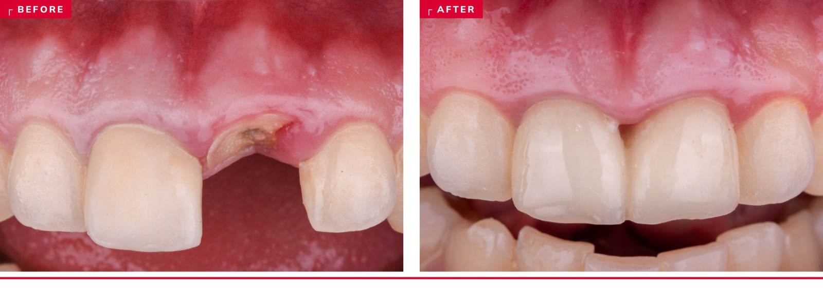
Immediate placement is the most demanding thing we do in the esthetic zone, and midfacial recession is how it fails. The instinct, when the tissue looks thin, is to reach for a graft. But margin stability isn't one move — it's a stack, and each tier can only protect what the one above it earned.
This issue assumes the foundation is already in place. Case selection and 3D position — Elian type, Kan sagittal root class, a palatal-biased shoulder, primary stability from apical and palatal bone, a fixture diameter chosen to preserve at least 2 mm of buccal gap rather than fill it — set the ceiling on everything below, and none of it is negotiable. A future issue will take that layer apart properly. What follows is what you do once the implant is in the right place: preserve what you're keeping, replace the deficit you couldn't prevent, and protect the result.
Tier 1 — Preserve
Don't destroy what you're keeping. The buccal plate is often under 1 mm and fed largely by the PDL — the cheapest bone is the bone you never lose.
How fragile the plate actually is. Chappuis and co-workers analyzed esthetic-zone sites with CBCT and found that at eight weeks post-extraction, a facial wall of ≤ 1 mm had lost a median of 7.5 mm of vertical buccal bone — against 1.1 mm where the wall was thicker (J Dent Res 2013). That is the most sobering number in this literature, and every decision in this tier follows from it.
Atraumatic extraction. Take the tooth out with that 7.5 mm in mind. Sever the attachment circumferentially, section multi-rooted teeth rather than luxating them as a unit, work vertically along the root, and never lever against the buccal. The forceps beak that feels efficient is a lever using a millimetre of bone as its fulcrum.
Stay flapless. Raising a buccal flap severs the periosteal supply on top of the PDL supply you just removed with the tooth — you cut the plate's blood supply twice in one appointment. Flapless placement preserved about 0.48 mm more horizontal buccal bone in Pitman and Cosyn's meta-analysis (p = .007), and the combination of flapless surgery, an intact wall, a thick phenotype and an immediate crown keeps advanced recession under 10% (Cosyn 2012).
The socket shield. This is the most elegant idea in the tier, and it works. Rather than extracting the buccal root fragment and then trying to rebuild what its removal cost you, you leave it in place — and with it, the plate's own periodontal attachment, its blood supply, and the bundle bone that would otherwise resorb on schedule. Nothing you graft afterward reproduces that. The outcomes bear it out: implant survival in the 96–99% range with measurably less bone loss (Oliva 2023; Kotsakis 2023), and 96.1% survival across Gluckman's 128-case series with up to four years of follow-up.
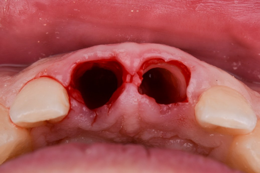
Managing a shield exposure. The technique's one recurring complication is internal shield exposure — the fragment migrating coronally or showing through the tissue — and the reason it shouldn't put you off is that it is genuinely simple to handle. You access the site, reduce and smooth the exposed portion, and let the tissue close over it; the implant stays. A multicenter series devoted specifically to this complication documents exactly that approach (Gluckman, Pohl & Chen 2024), and in the 128-case series the complications were likewise managed or observed without implant loss. Knowing what an exposure looks like and having a plan for it is the whole barrier to entry.
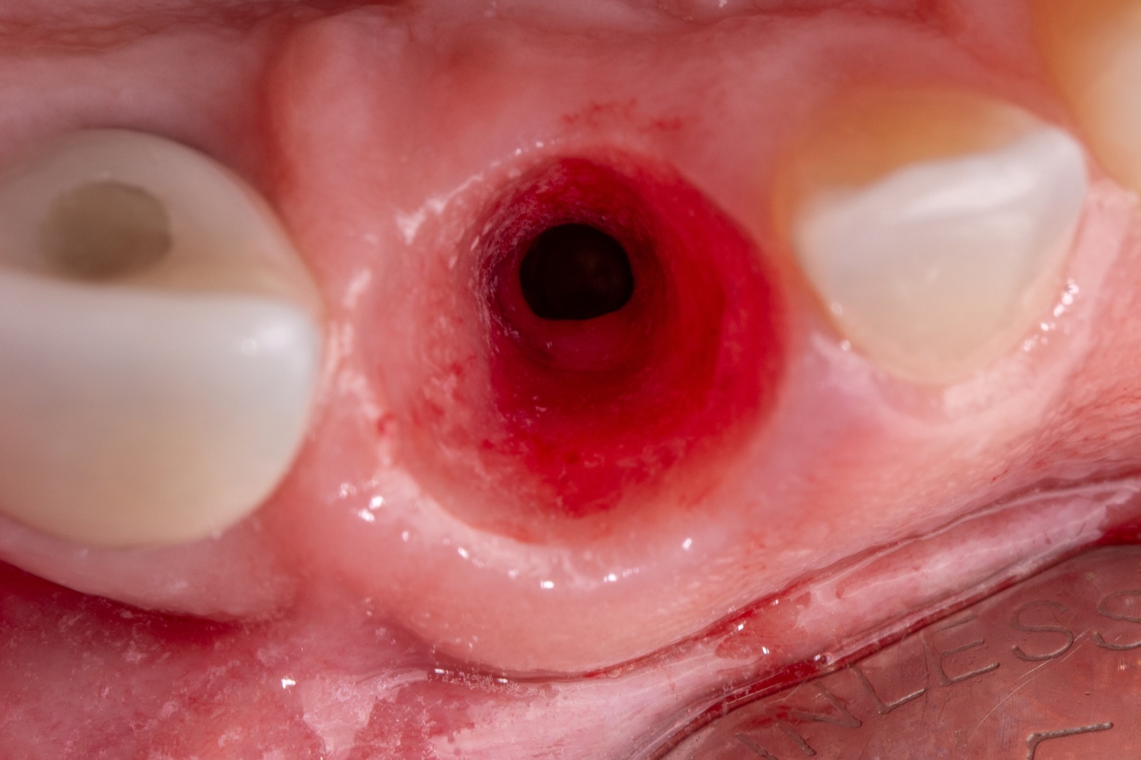
The proximal shield. The same logic extends interproximally: retain a proximal fragment to support the papilla between adjacent immediates, where papilla height is otherwise dictated by the bone on the neighbouring root. It's biologically sound and clinically satisfying, though the published support is still case-report level rather than controlled.
TIER 1, IN ONE LINE
Everything you preserve is bone and attachment you never have to rebuild — extract without leverage, leave the flap alone, and shield the plate when the case allows it.
Tier 2 — Replace
Rebuild the deficit you couldn't prevent — hard tissue first, then soft.
Graft the jumping gap. This is the best-evidenced hard-tissue move in the entire sequence. Seyssens' meta-analysis of 15 RCTs found that grafting the gap with a slow-resorbing xenograft cut horizontal buccal resorption by 0.59 mm (~54%) and midfacial soft-tissue migration by 0.58 mm, at GRADE moderate certainty. Araújo's CBCT work adds the dimensional case: grafted wide gaps (> 2 mm) preserved more than 90% of ridge dimension, while narrow gaps lost about 41%. There is no good argument for leaving the gap empty.
Add a connective tissue graft. Soft tissue is the other half of the margin. A simultaneous CTG shifts the midfacial level roughly 0.41 mm coronally and cuts the risk of ≥ 1 mm midfacial asymmetry about twelvefold (Seyssens 2021); in Zuiderveld's RCT, advanced recession fell from 32% to 8%. Deliver it flaplessly through a tunnel or VISTA approach — the entire point of Tier 1 was to protect that blood supply, so don't undo it here. Timing is a wash: simultaneous and staged grafting produce equivalent thickness (Lin 2018), so there's rarely a reason to make the patient come back.
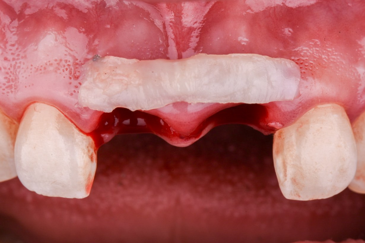
Dual zone, and the modified version. When a second surgical site isn't on the table, dual-zone grafting is the reasonable alternative. It matches CTG on esthetic scores (PES 12.1 vs 12.5, non-significant) but loses roughly twice the labial volume, and CTG holds contour better in thin-bone sites (Fettouh 2024 RCT). Carrying the particulate past the jumping gap and up into the tissue zone above the crest — the modified version — puts graft rather than clot underneath the transmucosal contour. Think of dual zone as the no-second-site option; CTG remains the volume play.
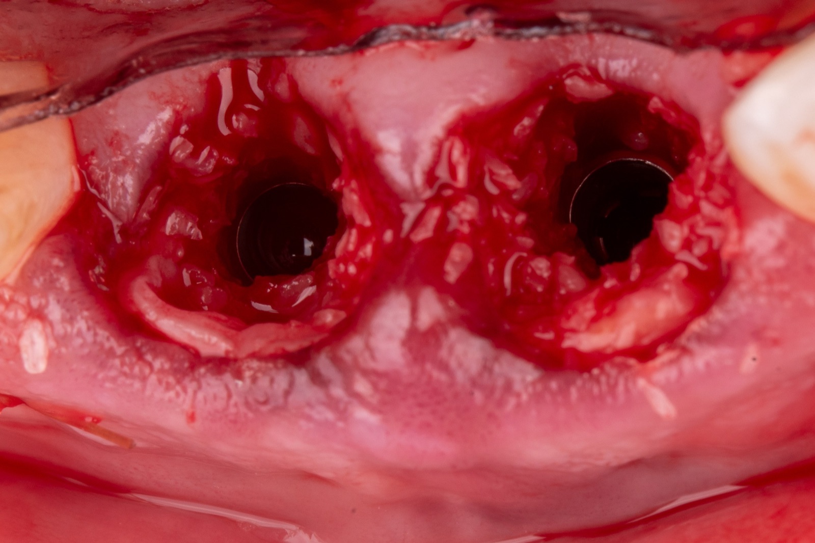
Substitutes, GBR, and biologics. Xenogeneic matrix and acellular dermal substitutes are non-inferior to CTG for thickness with less morbidity, but trend toward more contour loss at ten years (Strauss 2026) — CTG is still the reference standard when midfacial position is what you're protecting. Sockets with a frank buccal dehiscence can often still be managed at placement using internal and external xenograft with or without a membrane, holding buccal thickness at one year (Chen 2021), though that evidence is case-series level. As for biologics: L-PRF is a perfectly reasonable seal, but it has not independently improved buccal bone or margin position, and rhPDGF and EMD have essentially no controlled evidence in immediates. Use them if you like them; don't build a plan around them.
TIER 2, IN ONE LINE
Graft the gap every time, add a flaplessly-delivered CTG whenever the phenotype is thin or the margin matters, and treat everything else in this tier as a substitute rather than an equal.
Tier 3 — Protect
The provisional and its contour are the mold your healed tissue sets into.
Provisionalize immediately, out of occlusion. When selection is respected, immediate placement with immediate provisionalization performs beautifully — 99.2% survival at one year and 95.8% at five, with a net gain in pink esthetic score and midfacial recession that is small and non-significant (Wittneben 2023). The caveat is that selection genuinely is everything: immediates run about 4% lower survival than delayed placement overall (94.9% vs 98.9%, Cosyn 2019), and that gap is a case-selection artifact rather than a property of the technique. If primary stability is short, don't force it — a custom or socket-sealing healing abutment shapes emergence without loading.
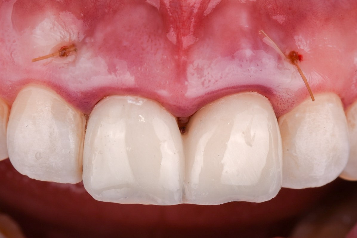
Contour: keep the subcritical zone concave. Su's critical/subcritical framework still governs the design. Keep the subcritical zone flat-to-concave so there is room for tissue volume, and make the critical contour a deliberate decision rather than whatever comes back from the lab.
Emergence angle only matters when the abutment is short. This is the part most people get wrong, and it's worth being precise. In our retrospective analysis of 192 implants in 119 patients, we split sites by transmucosal abutment height (TmAH) and restorative emergence angle (REA). The group with a short abutment (< 2 mm) and a wide angle (≥ 30°) — SW — lost significantly more marginal bone than every other group, and carried roughly four times the likelihood of a peri-implantitis diagnosis compared with the long-abutment groups. But the long-abutment groups did not differ from each other: with more than 2 mm of transmucosal height, a wide emergence angle stopped being a significant risk factor. Every additional millimetre of abutment height reduced the odds of any marginal bone loss (OR 0.63, p = .003). (Misch et al., Clin Oral Implants Res 2025.)
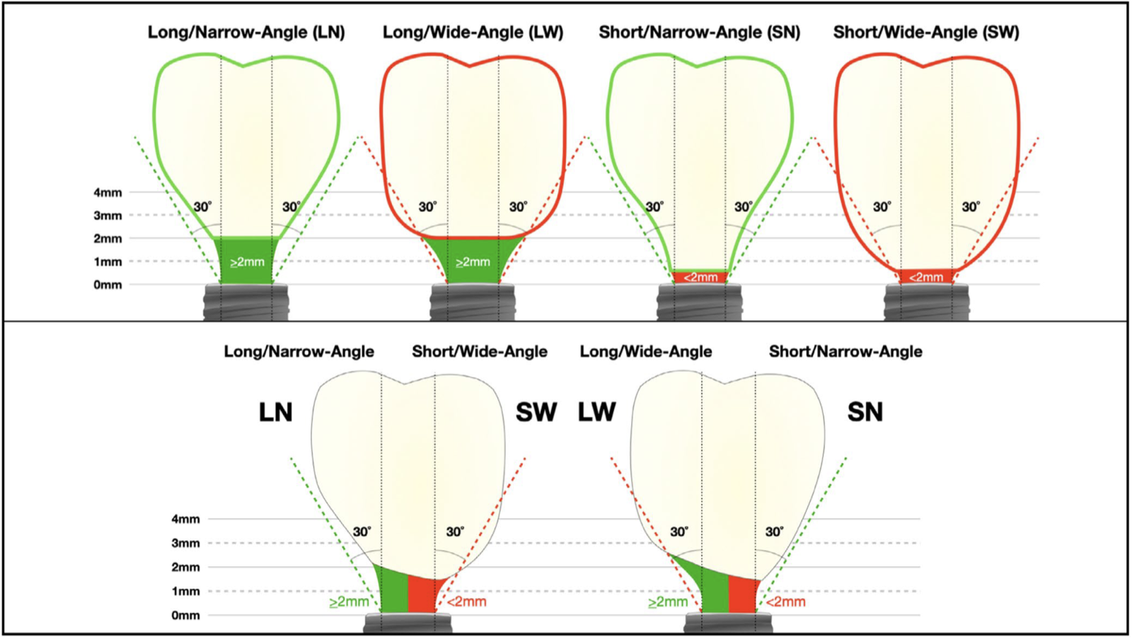
The practical translation is that emergence angle is not an independent villain — it is dangerous specifically when you have not given yourself vertical room. Buy that room where you can: place slightly subcrestal, choose a taller transmucosal component, and reserve your worry about a wide angle for the sites where you couldn't.
Plan for the access channel. Screw retention is preferable for all the usual reasons, but a straight access channel is anatomically possible only about 14% of the time in the anterior maxilla (Kan 2021). Decide how you're handling that before the provisional is made, not after.
TIER 3, IN ONE LINE
Provisionalize out of occlusion, keep the subcritical zone concave, and buy vertical abutment height — above 2 mm, emergence angle stops being the problem everyone treats it as.
START MONDAY
Five things to fold into your next immediate case:
- Extract like the plate is 1 mm thick, because it usually is. No buccal leverage, no flap.
- Shield when the case allows it — single-rooted, intact fragment — and learn to manage an exposure before your first one.
- Graft the gap every time, and add a flapless CTG when the phenotype is thin or the margin is precious.
- Provisionalize out of occlusion with a concave subcritical contour, or seal with a custom healing abutment if stability is short.
- Give yourself abutment height. Above 2 mm of transmucosal height, emergence angle stops being a significant risk.
THE HONEST VERSION
The soft-tissue moves in Tier 2 reliably protect the midfacial margin, but they cannot relocate a badly placed implant — which is why the foundation tier this issue skipped over is the one that actually sets your ceiling. The shield techniques do something no graft reproduces, and their one common complication is manageable, but the meta-analyses remain heterogeneous enough that confident numbers should be read with care. The biologics are, for now, unproven here. And the field's quiet caveat, flagged by Cosyn: midfacial recession is chronically under-measured against the true preoperative baseline — so read every "stable margin" claim with that in mind. Stack the tiers, respect case selection, and let grafts protect a good position rather than rescue a bad one.
MORE FROM MISCH IMPLANT IQ
All issues: Misch Implant IQ archive
UPCOMING MISCH INSTITUTE COURSES
Fresh dates are posted as they're announced — see the full institute calendar at misch.com.
Full calendar and registration at misch.com.
Sources
Chappuis V, Engel O, Reyes M, Shahim K, Nolte L-P, Buser D. Ridge alterations post-extraction in the esthetic zone: a 3D analysis with CBCT. J Dent Res. 2013;92(12 Suppl):195S–201S. · PubMed · DOI
Pitman J, Christiaens V, Callens J, et al. Immediate implant placement with flap or flapless surgery: a systematic review and meta-analysis. J Clin Periodontol. 2023;50(6):755–764. · PubMed · DOI
Cosyn J, Hooghe N, De Bruyn H. A systematic review on the frequency of advanced recession following single immediate implant treatment. J Clin Periodontol. 2012;39(6):582–589. · PubMed · DOI
Oliva S, Capogreco M, Murmura G, et al. The socket shield technique and its complications, implant survival rate, and clinical outcomes: a systematic review. J Periodontal Implant Sci. 2023;53(2):99–109. · PubMed · DOI
Kotsakis GA, Nguyen TT, Siormpas K, et al. Clinical outcomes of retention of the buccal root section combined with immediate implant placement: a systematic review. Clin Implant Dent Relat Res. 2023;25(1):23–34. · PubMed · DOI
Gluckman H, Salama M, Du Toit J. A retrospective evaluation of 128 socket-shield cases in the esthetic zone and posterior sites: partial extraction therapy with up to 4 years follow-up. Clin Implant Dent Relat Res. 2018;20(2):122–129. · PubMed · DOI
Gluckman H, Pohl S, Chen J. Socket shield complications: the management of internal shield exposure. A multicenter case series. Int J Periodontics Restorative Dent. 2024;44(2):177–185. · PubMed · DOI
Seyssens L, Eeckhout C, Cosyn J. Immediate implant placement with or without socket grafting: a systematic review and meta-analysis. Clin Implant Dent Relat Res. 2022;24(3):339–351. · PubMed · DOI
Araújo MG, Dias DR, Wang P, Levine RA. Effect of the buccal gap width on alveolar process reduction following immediate implant placement: a retrospective CBCT study. Clin Oral Implants Res. 2026;37(5):630–642. · PubMed · DOI
Seyssens L, De Lat L, Cosyn J. Immediate implant placement with or without connective tissue graft: a systematic review and meta-analysis. J Clin Periodontol. 2021;48(2):284–301. · PubMed · DOI
Zuiderveld EG, Meijer HJA, den Hartog L, Vissink A, Raghoebar GM. Effect of connective tissue grafting on peri-implant tissue in single immediate implant sites: a randomized controlled trial. J Clin Periodontol. 2018;45(2):253–264. · PubMed · DOI
Lin C-Y, Chen Z, Pan W-L, Wang H-L. Impact of timing on soft tissue augmentation during implant treatment: a systematic review and meta-analysis. Clin Oral Implants Res. 2018;29(5):508–521. · PubMed · DOI
Fettouh AIA, Ghallab NA, Mina NA, et al. Hard and soft tissue alterations using dual-zone concept versus connective tissue graft at maxillary immediate implant placement: a 1-year randomized clinical trial. Clin Oral Implants Res. 2024;35(5):510–525. · PubMed · DOI
Strauss FJ, Liguori MG, Gasser TJW, Naenni N, Jung RE, Thoma DS. Soft tissue volume augmentation at single implant sites applying collagen matrices or connective tissue grafts: 10-year follow-up of an RCT. J Esthet Restor Dent. 2026 (online). · PubMed · DOI
Chen K, Li Z, Liu X, et al. Immediate implant placement with buccal bone augmentation in the anterior maxilla with thin buccal plate: a one-year case series. J Prosthodont. 2021;30(6):473–480. · PubMed · DOI
Wittneben JG, Molinero-Mourelle P, Hamilton A, et al. Clinical performance of immediately placed and immediately loaded single implants in the esthetic zone: a systematic review and meta-analysis. Clin Oral Implants Res. 2023;34(Suppl 26):266–303. · PubMed · DOI
Cosyn J, De Lat L, Seyssens L, et al. The effectiveness of immediate implant placement for single tooth replacement compared to delayed placement: a systematic review and meta-analysis. J Clin Periodontol. 2019;46(Suppl 21):224–241. · PubMed · DOI
Su H, González-Martín O, Weisgold A, Lee E. Considerations of implant abutment and crown contour: critical contour and subcritical contour. Int J Periodontics Restorative Dent. 2010;30(4):335–343. · PubMed
Misch J, Abu-Reyal S, Lohana D, Mandil O, Saleh MHA, Li J, Wang H-L, Ravidà A. Combined effect of abutment height and restoration emergence angle on peri-implant bone loss progression: a retrospective analysis. Clin Oral Implants Res. 2025;36(5):600–612. · PubMed · DOI
Kan JYK, Rungcharassaeng K, Lin GH, et al. Incidence of straight-channel screw-retained single implant crown following immediate implant placement and provisionalization in the esthetic zone: a CBCT study. Int J Oral Maxillofac Implants. 2021;36(4):793–798. · PubMed · DOI
Elian N, Cho SC, Froum S, Smith RB, Tarnow DP. A simplified socket classification and repair technique. Pract Proced Aesthet Dent. 2007;19(2):99-104. · PubMed
Kan JY, Roe P, Rungcharassaeng K, et al.. Classification of sagittal root position in relation to the anterior maxillary osseous housing for immediate implant placement: a cone beam computed tomography study. Int J Oral Maxillofac Implants. 2011;26(4):873-876. · PubMed
Misch J, Alrmali AE, Galindo-Fernandez P, Saleh MHA, Wang HL. Peri-implant tissue stability: the PROS concept. Int J Oral Implantol (Berl). 2025;18(1):73-84. · PubMed
Misch Implant IQ · Educational content — not a substitute for individual clinical judgment. Figures are the most recent documented values from the cited studies.