

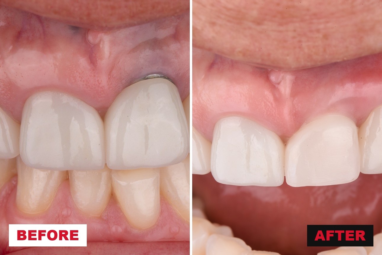
A patient points at a grey shadow above an anterior implant crown and asks what happened. The reflex is to book a connective tissue graft. That reflex is where most of these cases are lost — not because grafting is wrong, but because a coronally advanced flap has nothing to advance over when the crown itself is the obstacle. The restoration that helped create the deficiency is still sitting in the way, and no amount of surgical technique gets around it.
The literature is blunt about this. Coverage of peri-implant soft tissue dehiscences with a flap and graft alone has historically landed between 40% and 66% — and in Burkhardt's prospective cohort, where coverage decayed from 75% at one month to 66% at six, complete coverage was achieved in none of the sites (Burkhardt 2008; Anderson 2014). The same defects treated with a staged prosthetic-surgical protocol reach 96.3% mean coverage at one year and 99.2% at five, with complete coverage in 75% and 79% of sites respectively (Zucchelli 2013; Zucchelli 2018). The technique is not what changed. The order of operations did. This issue walks the staged sequence — diagnose, reshape, graft, re-restore — through a single case, and closes with the part nobody enjoys hearing: most of these are preventable at placement.
Stage 0 — Rule out disease, then classify
You are not allowed to call it esthetic until you have excluded peri-implantitis, and you cannot pick a technique until you know the class.
Disease first, and this is a hard gate. Every classification and decision tree in this space applies only to a clinically healthy implant. Following the 2017 World Workshop, an implant with bleeding on gentle probing, probing depth ≥ 6 mm, and bone levels ≥ 3 mm apical to the coronal portion of the intraosseous surface is diseased, not deficient (Berglundh 2018). Treating a peri-implantitis lesion with a bilaminar flap buries the problem under a graft. Probe, radiograph, and be honest about what you find before anything else happens.
Then classify by two things: where the crown sits, and how tall the papillae are. Zucchelli and co-workers built the reference system around the buccolingual position of the implant-supported crown, and then subdivided by papilla height (J Periodontol 2019). Class I is a margin in the right place with the grey showing through — a thickness problem, not a position problem. Class II is an apically positioned margin where the crown profile still sits inside the imaginary curve connecting the adjacent teeth. Classes III and IV are apically positioned margins where the crown profile sits outside that curve; the two are separated only by where the implant head itself lands once the crown is off, palatal (III) or facial (IV) to the line joining the adjacent gingival margins.
The subclass is what actually dictates the sequence. Subclass a means both papilla tips are ≥ 3 mm coronal to the ideal margin position; b means at least one is between 1 and 3 mm; c means at least one is under 1 mm. That single measurement decides whether you can advance a flap at all. Subclass a can go straight to CAF + CTG. Subclass b is the indication for the full prosthetic-surgical approach — you have to grow the papillae before you can borrow from them. Subclass c has no papillae to borrow from, so the site gets treated as edentulous: crown and abutment out, cover screw in, augment under submerged healing.
Alrmali's tree is the version to keep on the wall. Working from Michigan, Alrmali and colleagues distilled the same territory into four destinations — purely prosthetic, purely surgical, combined prosthetic-surgical (with or without abutment removal), and explantation — routed by three evaluations: three-dimensional implant position relative to the alveolar contour, hard tissue status, and soft tissue status, with keratinized mucosa width dichotomized at 2 mm (J Esthet Restor Dent 2023). It is the more practical instrument at the chair, because it forces the position question first. And it names the outcome we avoid saying out loud: when the implant head is frankly facial and the papillae are gone — Zucchelli's Class IVc — the honest answer is removal, not coverage.
STAGE 0, IN ONE LINE
Exclude disease, remove the crown to see the implant head, and let papilla height tell you whether you are grafting today or in two months.
Stage 1 — The prosthetic phase
Two months of deliberately under-contoured provisional is what buys you the tissue you are about to advance.
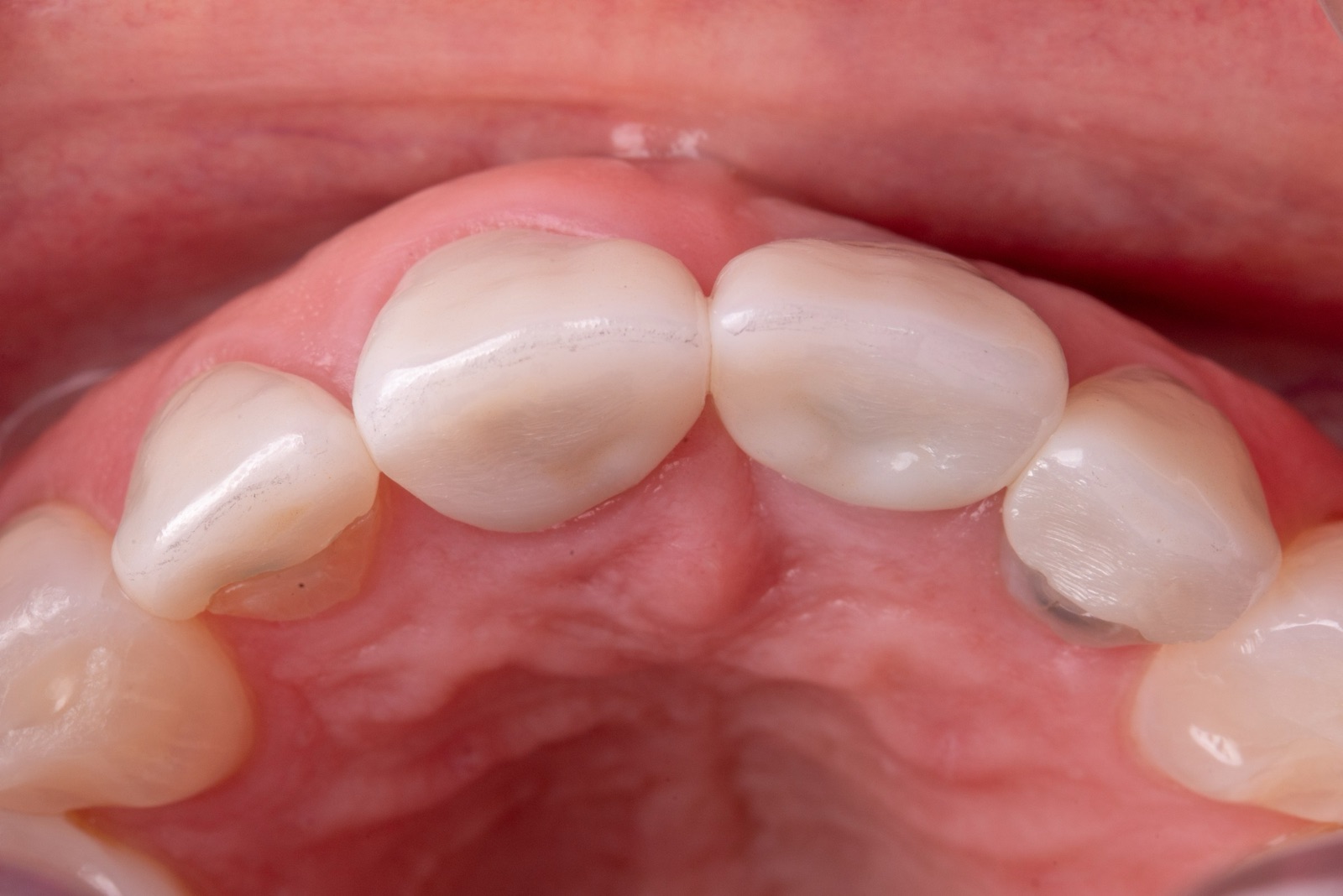
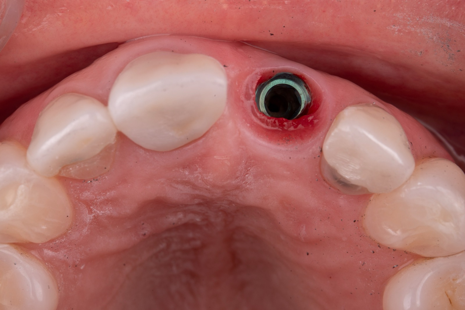
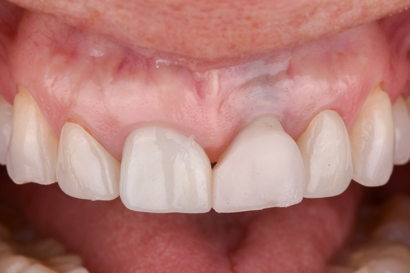
Take the crown off and shorten the abutment. In the case shown here — a maxillary left central with an apically positioned margin, visible metal at the collar, and a papilla that would not support advancement — the definitive crown came off, the abutment was reduced in height and hollowed, and a shortened screw-retained provisional went on with a deliberately flat-to-concave subgingival profile. The provisional is intentionally shorter than the final crown. You are not restoring esthetics in this phase; you are vacating space so tissue can move into it.
Then wait, because the papillae are growing. Zucchelli's protocol allows roughly two months between the provisional and the surgery, and the interproximal tissue responds by increasing in both height and thickness. This is the entire point. The coronally advanced flap in Stage 2 is fed and anchored by those papillae — they get de-epithelialized toward the palate and become the bed the flap sutures to. A flap advanced over shallow papillae has no blood supply and no anchorage, which is precisely why the crown-in-situ approach underperforms in these classes.
STAGE 1, IN ONE LINE
The provisional's job is to get out of the way for two months so the papillae can grow into the space the final crown will eventually occupy.
Stage 2 — The surgical phase
Coronally advanced flap plus a de-epithelialized connective tissue graft — and the graft is doing thickness work, not just coverage work.
Design the flap to be advanced, not just elevated. A trapezoidal envelope with split-full-split thickness elevation gives you the mobility to bring the margin above the abutment convexity and hold it there without tension. The anatomic papillae are de-epithelialized toward the palatal so the advanced flap has vascularized connective tissue to sit on rather than epithelium.
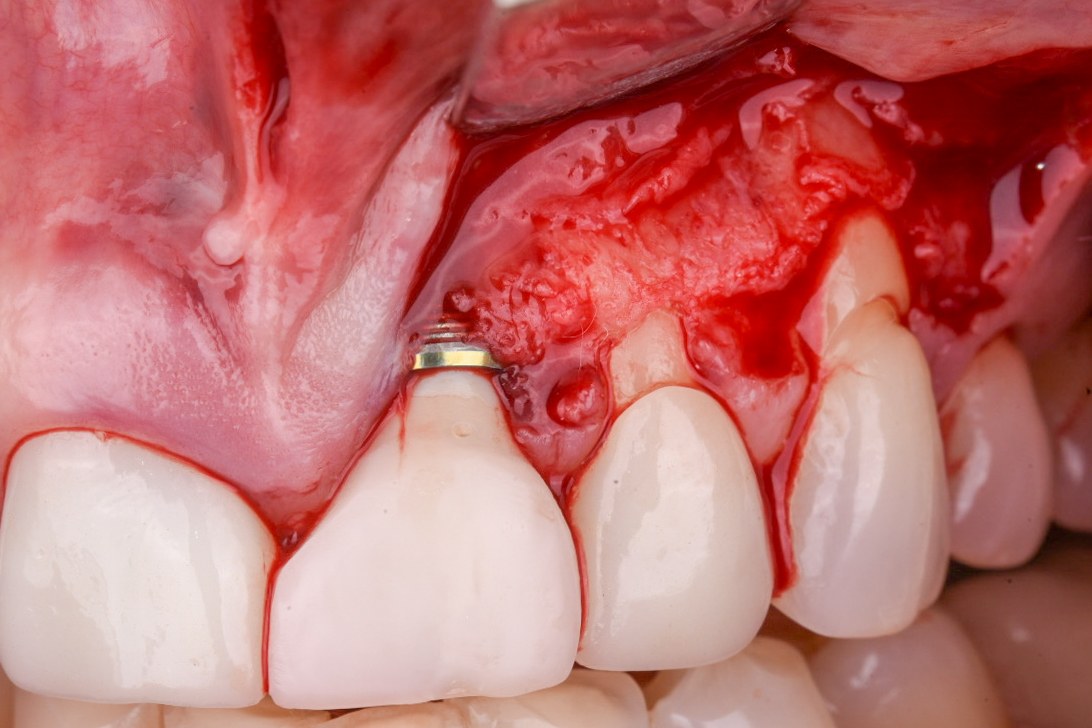
Use a de-epithelialized free gingival graft, not a subepithelial harvest. Zucchelli's group makes this distinction deliberately: a graft de-epithelialized from a free gingival graft is dense collagenous connective tissue with minimal fatty and glandular content, which makes it firmer, more stable, and better at holding volume than a subepithelial harvest (J Clin Periodontol 2010). Around implants, where the peri-implant connective tissue is scar-like — fibers parallel to the surface, fewer fibroblasts, reduced vascularity (Sculean 2017) — that quality difference matters more than it does on a root.
Fix the graft at the level of the contralateral tooth's margin. Not at the current margin, and not where the crown used to end. The reference is the homologous natural tooth, because that is the definition of a successful outcome in this classification. Thickness is the other target: Zucchelli's group puts the working threshold at roughly 2 mm of mucosal thickness before the grey of the abutment stops reading through the tissue (van Brakel 2011).
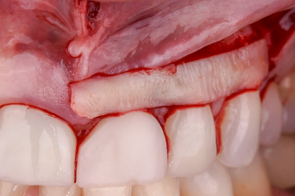
Close it passively, over the abutment. Primary closure above the abutment convexity is what protects the graft through the first weeks. If the flap needs tension to reach, the prosthetic phase was too short or the abutment was not reduced enough — go back rather than pulling.
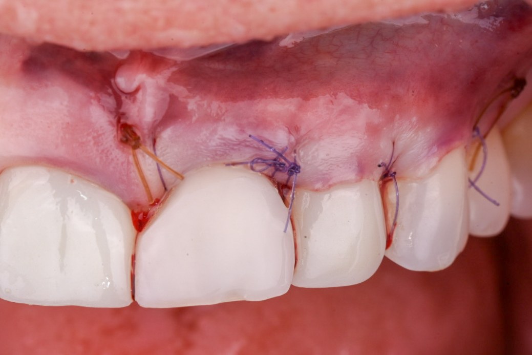
STAGE 2, IN ONE LINE
Advance to the contralateral margin, graft with de-epithelialized dense connective tissue for thickness, and close over the abutment without tension.
Stage 3 — Re-restoring, and the long tail
The margin keeps improving for years, which is exactly why you do not rush the definitive crown.
Condition first, restore late. After maturation, a new temporary crown conditions the augmented tissue, and the definitive restoration follows roughly 8 to 12 months after surgery. It feels slow. The reason to tolerate it is that the tissue is still moving. Buccal soft tissue thickness increased 1.54 ± 0.21 mm at one year, then gained a further 0.3 mm between years one and five (p < .001), with keratinized tissue height adding another 0.5 mm over the same window. Mean coverage climbed from 96.3% to 99.2% through creeping attachment (Eur J Oral Implantol 2018). A definitive crown delivered too early gets contoured against tissue that has not finished arriving.
Grafted sites hold. Across 15 prospective studies with at least three years of follow-up, CTG-based augmentation at implant sites produced stable or increasing keratinized mucosa width and mucosal thickness over time, stable marginal bone, and peri-implantitis incidence between 0% and 7.1% — while non-augmented implants tended to show continued apical migration of the margin (Stefanini 2023). The graft is not only cosmetic.
STAGE 3, IN ONE LINE
Condition, wait 8–12 months, and let creeping attachment finish the last few percent before you commit to a definitive contour.
Preventing the next one
Nearly half of all implants develop one of these, and most of the risk is set at placement.
The numbers are worse than the field's reputation suggests. The AO/AAP systematic review of 221 studies puts mean PSTD prevalence at 46.2%, with incidence up to 38.3% within five years of loading, in the absence of disease (Tavelli & Barootchi 2025). Restricted to single implants in the esthetic zone, prevalence measures 54.2% at the patient level (Tavelli 2022). This is not a rare complication we occasionally see; it is the default trajectory of an unprotected implant.
The risk factors are almost entirely ours. Limited mucosal thickness, immediate placement, and the absence of soft tissue augmentation at placement drive PSTD; guided surgery, bone grafting at placement, soft tissue augmentation, and adequate keratinized mucosa and thickness protect against it (Tavelli & Barootchi 2025). Add reduced keratinized mucosa width, longer time in function, and increased buccal bone distance from the ultrasonographic data (Tavelli 2022). Every item on the protective list is a decision made on the day of surgery.
Build the restorative half in from the start. The PROS framework — platform switching, restorative abutment design, an optimal internal conical connection, and subcrestal placement — organizes the prosthetic variables that keep peri-implant tissues stable rather than leaving them to be repaired later (Misch, Alrmali, Galindo-Fernandez, Saleh & Wang, Int J Oral Implantol 2025). Abutment height is the underrated member of that set: above 2 mm of transmucosal height, emergence angle largely stops behaving as an independent risk factor, and every additional millimetre of height lowers the odds of any marginal bone loss (Misch et al., Clin Oral Implants Res 2025).
PREVENTION, IN ONE LINE
Thickness at placement, a band of keratinized mucosa, guided position, and ≥ 2 mm of abutment height — the four things that keep you out of this issue entirely.
START MONDAY
Six things to fold into your next peri-implant soft tissue case:
- Probe and radiograph before you call it esthetic. BOP, PD ≥ 6 mm, or bone ≥ 3 mm apical to the intraosseous surface means you are treating disease, not a deficiency.
- Measure papilla height against the ideal margin, not the current one. ≥ 3 mm both sides means you can graft now; 1–3 mm means the crown comes off first.
- Take the crown off in any Class III or IV. You cannot classify the implant head — or advance a flap over the crown profile — with the restoration in place.
- Shorten the abutment and provisionalize for two months. Under-contour deliberately; the papillae you grow are the flap's blood supply.
- Harvest a de-epithelialized free gingival graft and fix it at the contralateral tooth's margin, aiming for ~2 mm of thickness.
- Wait 8–12 months for the definitive crown, and use the interval to condition rather than commit.
THE HONEST VERSION
The 96–99% coverage figures come from Zucchelli's own prospective cohorts, in his hands, with strict case selection — they are a ceiling, not an expectation. The classification itself is explicitly experience-based; the authors say plainly that it awaits validation. The subclass thresholds of 3 mm and 1 mm are clinically sensible but not empirically derived, and papilla measurement against an "ideal" margin position carries real inter-operator variability. Alrmali's tree shares that pedigree: a well-reasoned map built on case series rather than controlled comparisons. What is solidly evidenced is narrower and still worth acting on — soft tissue dehiscence is common, thin tissue and immediate placement without augmentation predict it, CTG-augmented sites stay stable for years, and removing the crown before advancing a flap changes the outcome by a margin no surgical refinement has matched. And the case shown here is one case: a good result in a Class III-type presentation with cooperative papillae, not proof of a technique.
MORE FROM MISCH IMPLANT IQ
All issues: Misch Implant IQ archive
UPCOMING MISCH INSTITUTE COURSES
Fresh dates are posted as they're announced — see the full institute calendar at misch.com.
Full calendar and registration at misch.com.
Sources
Berglundh T, Armitage G, Araujo MG, et al. Peri-implant diseases and conditions: consensus report of workgroup 4 of the 2017 World Workshop on the Classification of Periodontal and Peri-Implant Diseases and Conditions. J Clin Periodontol. 2018;45(Suppl 20):S286–S291. · PubMed · DOI
Zucchelli G, Tavelli L, Stefanini M, Barootchi S, Mazzotti C, Gori G, Wang H-L. Classification of facial peri-implant soft tissue dehiscence/deficiencies at single implant sites in the esthetic zone. J Periodontol. 2019;90(10):1116–1124. · PubMed · DOI
Alrmali A, Stuhr S, Saleh MHA, Latimer J, Kan J, Tarnow DP, Wang H-L. A decision-making tree for evaluating an esthetically compromised single dental implant. J Esthet Restor Dent. 2023;35(8):1239–1248. · PubMed · DOI
Zucchelli G, Mazzotti C, Mounssif I, Mele M, Stefanini M, Montebugnoli L. A novel surgical-prosthetic approach for soft tissue dehiscence coverage around single implant. Clin Oral Implants Res. 2013;24(9):957–962. · PubMed · DOI
Zucchelli G, Felice P, Mazzotti C, et al. 5-year outcomes after coverage of soft tissue dehiscence around single implants: a prospective cohort study. Eur J Oral Implantol. 2018;11(2):215–224. · PubMed
Mazzotti C, Stefanini M, Felice P, Bentivogli V, Mounssif I, Zucchelli G. Soft-tissue dehiscence coverage at peri-implant sites. Periodontol 2000. 2018;77(1):256–272. · PubMed · DOI
Burkhardt R, Joss A, Lang NP. Soft tissue dehiscence coverage around endosseous implants: a prospective cohort study. Clin Oral Implants Res. 2008;19(5):451–457. · PubMed · DOI
Anderson LE, Inglehart MR, El-Kholy K, Eber R, Wang H-L. Implant associated soft tissue defects in the anterior maxilla: a randomized control trial comparing subepithelial connective tissue graft and acellular dermal matrix allograft. Implant Dent. 2014;23(4):416–425. · PubMed · DOI
Zucchelli G, Mele M, Stefanini M, Mazzotti C, Marzadori M, Montebugnoli L, De Sanctis M. Patient morbidity and root coverage outcome after subepithelial connective tissue and de-epithelialized grafts: a comparative randomized-controlled clinical trial. J Clin Periodontol. 2010;37(8):728–738. · PubMed · DOI
Sculean A, Chappuis V, Cosgarea R. Coverage of mucosal recessions at dental implants. Periodontol 2000. 2017;73(1):134–140. · PubMed · DOI
van Brakel R, Noordmans HJ, Frenken J, de Roode R, de Wit GC, Cune MS. The effect of zirconia and titanium implant abutments on light reflection of the supporting soft tissues. Clin Oral Implants Res. 2011;22(10):1172–1178. · PubMed · DOI
Stefanini M, Barootchi S, Sangiorgi M, et al. Do soft tissue augmentation techniques provide stable and favorable peri-implant conditions in the medium and long term? A systematic review. Clin Oral Implants Res. 2023;34(Suppl 26):28–42. · PubMed · DOI
Tavelli L, Barootchi S. Prevalence, incidence, risk, and protective factors for soft tissue dehiscences at implant sites in the absence of disease: an AO/AAP systematic review and meta-regression analysis. J Periodontol. 2025;96(6):562–586. · PubMed · DOI
Tavelli L, Barootchi S, Majzoub J, et al. Prevalence and risk indicators of midfacial peri-implant soft tissue dehiscence at single site in the esthetic zone: a cross-sectional clinical and ultrasonographic study. J Periodontol. 2022;93(6):857–866. · PubMed · DOI
Misch J, Alrmali AE, Galindo-Fernandez P, Saleh MHA, Wang H-L. Peri-implant tissue stability: the PROS concept. Int J Oral Implantol. 2025;18(1):73–84. · PubMed
Misch J, Abu-Reyal S, Lohana D, Mandil O, Saleh MHA, Li J, Wang H-L, Ravidà A. Combined effect of abutment height and restoration emergence angle on peri-implant bone loss progression: a retrospective analysis. Clin Oral Implants Res. 2025;36(5):600–612. · PubMed · DOI
Misch Implant IQ · Educational content — not a substitute for individual clinical judgment. Figures are the most recent documented values from the cited studies.